Новини ринку

Radiography as one of the methods of radiologic diagnosis of coronavirus disease (COVID-19)

Principles of selection of radial methods of research:
  1. Symptoms and clinical signs of ARVI are absent (even against the background of relevant anamnestic data) – the use of radiation studies is not indicated.
  2. Symptoms and clinical signs of acute respiratory infection are present – OHC radiography is recommended.
  3. Symptoms and clinical signs of acute respiratory infections are present, there is a suspicion of COVID-19 (including on the basis of anamnestic data) – computed tomography of the OHC is recommended.
  4. Evaluation of the dynamics of verified viral pneumonia caused by 2019-nCoV (COVID-19):
  • CT scan of the OHC;
  • OGC radiography and/or OGC ultrasound (additional method) in patients in critical condition, in intensive care and intensive care units, if transportation is impossible or if CT scanning is not possible.
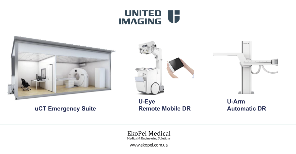
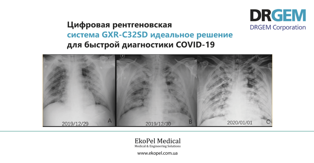
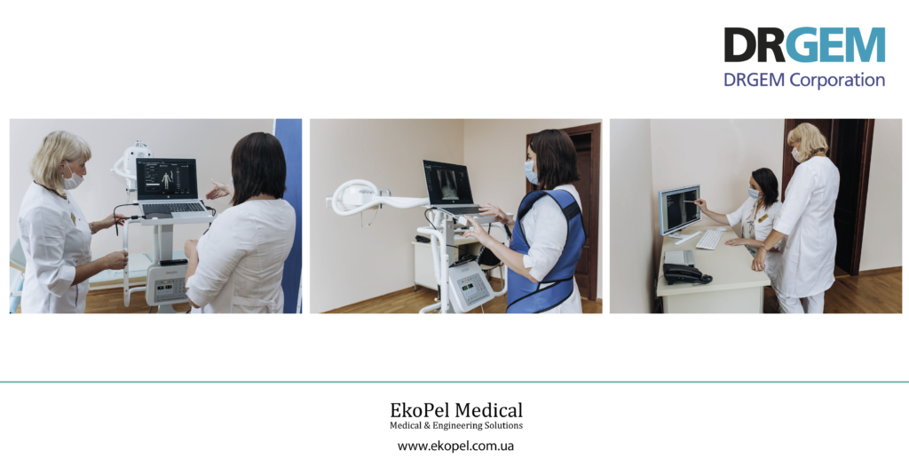
OGC RADIOGRAPHY IS USED IN OUTPATIENT AND INPATIENT SETTINGS AS PART OF THE SCREENING PROGRAM FOR SUSPECTED CORONAVIRUS INFECTION. The appointment of this study is made based on the peculiarities of symptoms and clinical manifestations of acute respiratory infections. Based on the results of chest radiography (including suspicion of COVID-19), the patient may be routed in accordance with current regulations or referred for additional examination by computed tomography of the chest. Inpatient radiography (RG) is recommended for use in patients in critical condition, in intensive care units and intensive care units, when it is impossible to transport them. In such cases, OGC X-ray is performed with a mobile X-ray machine (including daily, on a scheduled basis). SEMIOTICS AND DIFFERENTIAL DIAGNOSIS The COVID-19 diagnostic algorithm is represented by a combined evaluation of epidemiologic history, clinical presentation, radiology and laboratory findings. RENTGENOGRAPHY OGC radiography is performed in anterior direct and lateral* 3 projections. When performing OHC X-ray, the main radiologic manifestations of viral pneumonia (including COVID-19) include: – numerous “frosted glass” type thickenings of rounded shape and varying extent (more often multilobar peripheral or basal location); – confluent infiltrative lesions; – thickening of lung tissue of alveolar type. More often the lesion is bilateral in nature. The volume of involvement of lung segments correlates with the severity of the disease course (Fig.1). Manifestations on OGC RG that are atypical for COVID-19: unilateral “frosted glass” type lung tissue thickening of central and root localization, pleural effusion, infiltration, cavitation, lymphadenopathy. It should be remembered that radiotherapy methods are not the main methods in the diagnosis of coronavirus infection, being characterized by high sensitivity but low specificity. The only specific diagnostic method is reverse transcription polymerase chain reaction (RT-PCR). Even if radiologic findings (computed tomography, radiography) of the thoracic organs (chest X-ray) are sufficiently suggestive of viral pneumonia, of which COVID-19 pneumonia is a variant, verification by laboratory tests (RT-PCR) and consistency with the clinical and laboratory picture are still required. * If the localization of the inflammatory process is unknown, it is advisable to perform a right lateral view. ** Image sources: Silverstein W. K., Stroud L., Cleghorn G. E., Leis J. A. First imported case of 2019 novel coronavirus in Canada presenting as mild pneumonia // Lancet. – 2020. – Feb. 29. – Vol. 395(10225):734. – Doi: 10.1016/S0140-6736(20)30370-6; Yoon S. H., Lee K. H., Kim J. Y., et al. Chest Radiographic and CT Findings of the 2019 Novel Coronavirus Disease (COVID-19): Analysis of Nine Patients Treated in Korea // Korean J Radiol. – 2020. – Apr. – Vol. 21, №4. – P. 494-500. – Doi: 10.3348/kjr.2020.0132.

Related Posts

Leave a Reply

Your email address will not be published. Required fields are marked *